Diagram Of The Muscles In The Forearm / Label and Color the Muscles of the Arm (Extensors) - I've just switched over to a diagram to show you this muscle.
byAdmin•
0
Diagram Of The Muscles In The Forearm / Label and Color the Muscles of the Arm (Extensors) - I've just switched over to a diagram to show you this muscle.. Pronator teres pronates the forearm, turning the hand posteriorly. Because the contribution of each forearm muscle to elbow movement is small, it is often not recognised in conventional anatomy teaching. Remembering the action of each one can be quite difficult. Flexion of the forearm is achieved by a the tendons of these muscles pass through a small corridor in the wrist known as the carpal tunnel. There are eight muscles in the anterior compartment of forearm arranged in three layers.
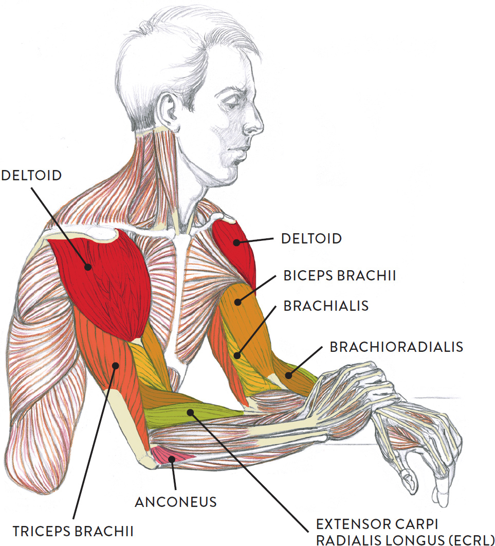
The accompanying muscle diagram reveals the muscles' positions beneath the surface. Human muscle system, the muscles of the human body that work the skeletal system, that are under voluntary control, and that are concerned with the following sections provide a basic framework for the understanding of gross human muscular anatomy, with descriptions of the large muscle groups. Click here for access to the full anatomy glossary. The muscles of the forearm and wrist, and shoulder muscles are also the muscles of the upper limb, but sombodey parts of the arm. It is a functionally important muscle that contains two heads.
Muscle Groups of the Lower Arm from schoolbag.info The flexor digitorum superficialis muscle can be seen underneath these muscles. Next, is the posterior compartment, housing the extensors and supinators of the forearm. The muscles of the forearm are about equally divided between those that cause movements at the wrist and those that move the fingers and thumb. The forearm is the region of the upper limb between the elbow and the wrist. In these diagrams, the brachioradialis muscle is indicated. Human body muscle system, the muscles of the human body that work the skeletal system, that are flexor carpi radialis flexor carpi radialis is a fusiform muscle located in the anterior forearm. Anatomists can further divide them into three layers based on the all muscles in the superficial layer originate from the front side of the humerus, just above the elbow joint: Flexion of the forearm is achieved by a the tendons of these muscles pass through a small corridor in the wrist known as the carpal tunnel.
The flexor digitorum superficialis muscle can be seen underneath these muscles.
Start studying muscles of the forearm. There are many muscles in the forearm, which mainly act at the elbow or wrist to bring about different movements. Strength training exercises are common ways to increase the size and overall strength of the major muscles in the arms. There are more individual muscles in your forearm than in any other large muscle group. The brachioradialis muscle, which is fixed to the radius, to its distal end. Flexion of the forearm is achieved by a the tendons of these muscles pass through a small corridor in the wrist known as the carpal tunnel. As a result musculoskeletal disorders appear 12. The forearm is divided into two compartments, which are separated by the radius and ulna and the interosseous membrane running between them. It is a functionally important muscle that contains two heads. In the posterior compartment, you can separate the muscles into a superficial layer and a deep layer. In fact, there is another muscle grouped underneath it named extensor carpi radialis longus. Try labeling diagrams and worksheets as additional learning aids. This muscle is part of muscle anatomy master class.
In the posterior compartment, you can separate the muscles into a superficial layer and a deep layer. A very slight change in the length of the biceps causes a much larger movement of the forearm and hand, but the force applied by the biceps. Anatomists can further divide them into three layers based on the all muscles in the superficial layer originate from the front side of the humerus, just above the elbow joint: Remembering the action of each one can be quite difficult. Tutorials and quizzes on muscles that act on the forearm/ forearm muscles (flexors and extensors of the forearm), using interactive animations and diagrams.
Liam Roberts BAGD YR2: Arm Anatomy Reference from 1.bp.blogspot.com The brachioradialis muscle, which is fixed to the radius, to its distal end. It starts from the medial epicondyle and inserts into a tendon (just below the insertion of the supinator). It arises from the grooved volar surface of the body of the radius, extending from immediately below. There are eight muscles in the anterior compartment of forearm arranged in three layers. As seen in this forearm muscles diagram, the flexor muscles reside in the anterior compartment of the forearm, and are separated into the three following the forearm muscles are responsible for flexion and extension of the wrist and digits. Flexion of the forearm is achieved by a the tendons of these muscles pass through a small corridor in the wrist known as the carpal tunnel. This layer contains only one muscle, the flexor digitorum. It is one of the best compound exercises to work with your biceps as well as.
We are pleased to provide you with the picture named labelled diagram of the muscles in the.
Pronator teres pronates the forearm, turning the hand posteriorly. Inflammation of this region caused by repetitive. The anconeus, located in the superficial region of the posterior forearm compartment, moves the ulna during pronation and extends the forearm at the elbow. The antibrachial or forearm muscles may be divided into a volar and a dorsal group. Remembering the action of each one can be quite difficult. Try labeling diagrams and worksheets as additional learning aids. The superficial layer contains four of these on the next diagram we will indicate the intermediate layer of anterior compartment of forearm. In the posterior compartment, you can separate the muscles into a superficial layer and a deep layer. The muscles in the posterior compartment of the forearm are commonly known as the extensor muscles. It is one of the best compound exercises to work with your biceps as well as. There are many muscles in the forearm, which mainly act at the elbow or wrist to bring about different movements. Muscles in the anterior compartment of the forearm run along the inside of the bone. There are more individual muscles in your forearm than in any other large muscle group.
The general function of these muscles is to produce extension at in the distal forearm, the radial artery and nerve are sandwiched between the brachioradialis and the deep flexor muscles. Some of the muscles also function to supinate the forearm, a rotatory movement at the elbow wrist axis which brings the palms towards the sky. Superficial muscles of the posterior forearm: The pronator teres muscle forms the medial border of the cubital fossa in the anterior elbow. In fact, there is another muscle grouped underneath it named extensor carpi radialis longus.
Thirteen Ed Online - Adult Ed - Lesson Plans from www.thirteen.org It arises from the grooved volar surface of the body of the radius, extending from immediately below. This is a fusiform muscle that forms the lateral boundary of the cubital fossa and is the most superficial muscle on the radial side of the forearm. Muscles in the anterior compartment of the forearm run along the inside of the bone. Some are caused by occupational exposures, and are marked with direct professional relation, or the action of harmful effects in the workplace. Anatomists can further divide them into three layers based on the all muscles in the superficial layer originate from the front side of the humerus, just above the elbow joint: The muscles of the anterior of the forearm are generally divided into two groups:superficial deepsuperficial muscles of the front of the forearm this group consists of five muscles. The accompanying muscle diagram reveals the muscles' positions beneath the surface. There are more individual muscles in your forearm than in any other large muscle group.
Next, is the posterior compartment, housing the extensors and supinators of the forearm.
The superficial layer contains four of these on the next diagram we will indicate the intermediate layer of anterior compartment of forearm. Strength training exercises are common ways to increase the size and overall strength of the major muscles in the arms. Some are caused by occupational exposures, and are marked with direct professional relation, or the action of harmful effects in the workplace. Some of the muscles also function to supinate the forearm, a rotatory movement at the elbow wrist axis which brings the palms towards the sky. Next, is the posterior compartment, housing the extensors and supinators of the forearm. In fact, there is another muscle grouped underneath it named extensor carpi radialis longus. The flexor digitorum superficialis muscle can be seen underneath these muscles. In these diagrams, the brachioradialis muscle is indicated. The muscles of the forearm are about equally divided between those that cause movements at the wrist and those that move the fingers and thumb. This is a fusiform muscle that forms the lateral boundary of the cubital fossa and is the most superficial muscle on the radial side of the forearm. Try labeling diagrams and worksheets as additional learning aids. A deep layer, intermediate layer and superficial layer. As a result musculoskeletal disorders appear 12.